Original Research | Published: 4 November 2022
open-url

Assessing the development status of intraoperative fluorescence imaging for anatomy visualisation, using the IDEAL framework

https://doi.org/10.1136/bmjsit-2022-000156

Request reuse permissionopen-url
This is an open access article distributed in accordance with the Creative Commons Attribution Non Commercial (CC BY-NC 4.0) licenseopen-url

Abstract

Objectives Intraoperative fluorescence imaging is currently used in a variety of surgical fields for four main purposes: visualising anatomy, assessing tissue perfusion, identifying/localising cancer and mapping lymphatic systems. To establish evidence-based guidance for research and practice, understanding the state of research on fluorescence imaging in different surgical fields is needed. We evaluated the evidence on fluorescence imaging used to visualise anatomical structures using the IDEAL framework, a framework designed to describe the stages of innovation in surgery and other interventional procedures.

Design IDEAL staging based on a thorough literature review.

Setting All publications on intraoperative fluorescence imaging for visualising anatomical structures reported in PubMed through 2020 were identified for five surgical procedures: cholangiography, hepatic segmentation, lung segmentation, ureterography and parathyroid identification.

Main outcome measures The IDEAL stage of research evidence was determined for each of the five procedures using a previously described approach.

Results 225 articles (8427 cases) were selected for analysis. Current status of research evidence on fluorescence imaging was rated IDEAL stage 2a for ureterography and lung segmentation, IDEAL 2b for hepatic segmentation and IDEAL stage 3 for cholangiography and parathyroid identification. Enhanced tissue identification rates using fluorescence imaging relative to conventional white-light imaging have been documented for all five procedures by comparative studies including randomised controlled trials for cholangiography and parathyroid identification. Advantages of anatomy visualisation with fluorescence imaging for improving short-term and long-term postoperative outcomes also were demonstrated, especially for hepatobiliary surgery and (para)thyroidectomy. No adverse reactions associated with fluorescent agents were reported.

Conclusions Intraoperative fluorescence imaging can be used safely to enhance the identification of anatomical structures, which may lead to improved postoperative outcomes. Overviewing current research knowledge using the IDEAL framework aids in designing further studies to develop fluorescence imaging techniques into an essential intraoperative navigation tool in each surgical field.

What is already known on this topic

  • Intraoperative fluorescence imaging has become widely used for identifying anatomical structures.

What this study adds

  • Based on the literature reviews, the IDEAL stage of fluorescence imaging for anatomy visualisation was determined for the following five surgical procedures: cholangiography (stage 3), hepatic segmentation (2b), lung segmentation (2a), ureterography (2a) and parathyroid identification (3).

How this study might affect research, practice or policy

  • Identifying the IDEAL stage of current research knowledge can aid in planning further studies to develop fluorescence imaging into an essential intraoperative navigational tool.

Introduction

Over the last two decades, intraoperative fluorescence imaging has become widely used to visualise biological structures, physiological function and neoplastic transformation more clearly than naked-eye inspections or conventional white-light examinations.1 2 In 2014, the International Society for Fluorescence-Guided Surgery (ISFGS) was established to further develop and disseminate fluorescence imaging techniques by sharing information among surgeons, researchers and industry engineers beyond specialties.3 During discussions at the sixth Annual Meeting of the ISFGS in 2019, we launched a project to overview the development of intraoperative fluorescence imaging across surgical fields and applications, using the IDEAL framework4 5 to identify which indications for fluorescence-guided surgery were ready for randomised controlled trials (RCTs) and which preparatory studies were needed in the other fields to increase the feasibility and value of future RCTs. The IDEAL framework describes five stages—Idea, Development, Exploration, Assessment and Long-term follow-up—in the evaluation of the life-cycle of surgical techniques, which can be recognised for a given technique by the types of study already published in which the technique was assessed.6

In this project, fluorescence imaging applications were classified into (1) perfusion assessment, (2) anatomy visualisation, (3) cancer identification and (4) lymphatic system identification. Among them, the use of fluorescence imaging for perfusion assessments has already been assessed successfully, employing the IDEAL framework (figure 1), after reviewing 196 articles published from 2002 to 2019.7 During this prior analysis, the current status of research evidence on perfusion assessments with fluorescence imaging was determined to be IDEAL stage 2a for upper gastrointestinal surgery and solid-organ transplantation, IDEAL 2b for coronary artery bypass grafting and IDEAL stage 3 for reconstructive surgery. The aim of the present study was to evaluate the current development status of fluorescence imaging for the intraoperative identification of anatomical structures in the same manner.

Figure 1
Figure 1

Flowchart of the IDEAL staging system. RCT, randomised controlled trial. Cited from a webpage of the IDEAL Collaboration: http://www.ideal-collaboration.net/ideal-flowchart-and-stage-questions-for-assessing-literature/

The main objective of using intraoperative fluorescence imaging for visualising anatomical structures is to decrease the risk of postoperative complications due to injuries or functional deficits affecting remaining organs. Another expected role of this technique is to guide resection of solid organs along accurate anatomical boundaries, which can enhance cancer curability by eradicating possible extension from the main tumour in the corresponding visceral segment. In this study, current knowledge was assessed on (a) the impact of anatomy visualisation by fluorescence imaging on these operative factors and (b) the method by which establishment and tissue identifiability. This was accomplished following the IDEAL framework, after reviewing previous publications focusing on the following five major applications to visualise bile ducts (cholangiography); segmental boundaries of the liver (hepatic segmentation) and lung (lung segmentation); ureters (ureterography) and parathyroids (thyroidectomy/ parathyroidectomy).

Methods

A literature review was conducted, through 31 December 2020, of all English-language publications in PubMed and Ovid/Medline reporting the techniques and outcomes of intraoperative fluorescence imaging used to visualise anatomical structures. The following keywords were used for both the title and abstract fields to identify potentially pertinent studies:

Cholangiography: (“fluorescence” OR “fluorescent” OR “near infrared imaging” OR “indocyanine green” OR “ICG”) AND (“cholecystectomy” OR “bile duct” OR “biliary tract” OR “cholangiography” OR “cholangiogram” OR “Calot’s triangle”)

Hepatic segmentation: (“fluorescence imaging” OR “fluorescent imaging” OR “near infrared imaging” OR “indocyanine green” OR “ICG”) AND (“hepatectomy” OR “liver resection” OR “hepatic segment” OR “liver segment”)

Lung segmentation: (“fluorescence imaging” OR “fluorescent imaging” OR “near infrared imaging” OR “indocyanine green” OR “ICG”) AND (“lung surgery” OR “lung resection” OR “lung segment” OR “pulmonary surgery” OR “thoracoscopic segmentectomy” OR “thoracoscopy”)

Ureterography: (fluorescence imaging” OR “fluorescent imaging” OR “near infrared imaging” OR “indocyanine green” OR “ICG”) AND (“ureter” OR “ureterography” OR “urinary tract” OR “pelvic surgery” OR “gynecological surgery” OR “urological surgery” OR “rectal surgery”)

Parathyroid identification: (“fluorescence imaging” OR “fluorescent imaging” OR “near infrared imaging” OR “indocyanine green” OR “ICG”) AND (“parathyroid” OR “thyroid” OR “accessory thyroid” OR “thyroidectomy”)

Inclusion criteria

Reports had to provide enough detail on fluorescence-guided surgery to allow readers to potentially reproduce the technique, and to report either technical or clinical (or both) outcomes of clinical use.

Exclusion criteria

Non-human preclinical studies, meta-analyses/systemic reviews, supplementary articles and conference proceedings were excluded. We also excluded articles with a focus outside the stated target application and those with inadequate information on patients’ background, treatment and/or outcomes to permit evaluation of what was done to whom and which outcomes were assessed.

Following the initial screening, each report’s adequacy was judged by an expert panel (figure 2).

Figure 2
Figure 2

Time and regional trends for publications on intraoperative fluorescence imaging for visualising anatomical structures. Time and regional trends for publications on intraoperative fluorescence imaging for anatomy visualisation were summarised for the five surgical procedures.

Figure 3
Figure 3

Time and regional trends for reported cases of intraoperative fluorescence imaging for visualising anatomical structures. Time and regional trends for reported cases on intraoperative fluorescence imaging for anatomy visualisation were summarised for the five surgical procedures.

Analysis

The maturity stage of intraoperative fluorescence imaging for anatomical identification was determined using the IDEAL framework (table 1).4–6 Since the reports studied were not written in IDEAL style, the stage of the research field was determined by allocating studies to IDEAL stages retrospectively, using the following heuristic: reports of only one or very few cases were classified as IDEAL stage 1 (idea). Outcomes of fluorescence imaging reported from case series and cohort studies were classified as IDEAL stage 2a (Development) or stage 2b (Exploration), with reports involving only a few centres (five or fewer) and with case series of <100 cases classed as stage 2a and larger studies as stage 2b. Fluorescence imaging techniques that had been evaluated in one or more multicentre RCTs were considered stage 3 (Assessment) and long-term evaluations using registries or databases classified as stage 4 (long term). The IDEAL stage proposed by the expert panels was determined after consensus formation in the observer panel about the overall state of the research field (online supplemental figure 1), rather than assigning the highest stage possible based on single studies.

Table 1
|
Breakdown of key considerations used to determine IDEAL stage

Results

Overview

The research keywords intended to include all reports related to intraoperative fluorescence imaging for anatomy visualisation identified 525 articles for initial screening. After applying the inclusion and exclusion criteria, 225 articles, reporting on a total of 8427 patients, were submitted for further evaluation (online supplemental figure 1).

Numbers of previous publications and numbers of reported cases are summarised, by year and by regional origin (Asia, Europe, North America, South America, Middle East, Oceania), in figures 2 and 3, respectively. The use of intraoperative fluorescence imaging for anatomy visualisation was first reported in the field of hepatobiliary surgery in 2008, followed by lung segmentation and parathyroid identification. Techniques and outcomes of hepatic and lung segmentation have been reported mainly from Asian regions, while publications from Western countries are dominant in the field of ureterography and parathyroid identification. Use of fluorescence imaging for cholangiography, which was initially introduced by Japanese surgeons, has recently been reported worldwide after publications of large case series and multi-institutional prospective studies from the USA, European countries and Argentina.

Summary of surveillance for each surgical field

Results on the literature surveillance for each surgical field are summarised, in terms of study design and imaging techniques in table 2, while endpoint measures and additional information used to determine each field’s IDEAL stage are summarised in table 3. Background data for each article are summarised in online supplemental tables 1–5. For all five surgical procedures, indocyanine green (ICG) was the main fluorescence probe used, while methylene blue (MB) and autofluorescence also were used for ureterography and parathyroid identification, respectively, with no reporting on adverse effects.

Table 2
|
Summary of previous publications on intraoperative fluorescence imaging for anatomy visualisation, in terms of study design and imaging techniques
Table 3
|
Summary of previous publications on intraoperative fluorescence imaging for anatomy visualisation, in terms of endpoint measures and additional information

Cholangiography

For cholangiography, our search yielded 68 eligible reports, 51 (75%) of them case series with no control arm, involving five or fewer centres and under 100 patients. There was one large multicentre prospective study8 and three RCTs.8–10 In total, 3386 cases were reported, the maximum number of patients in any study being 676 (online supplemental table 1).

Intravenously injected ICG at either an absolute dose of 2.5 mg per patient and given 15–120 min prior to surgery was used in 44% of studies, while some authors reported intrabiliary injection of ICG in concentrations ranging from 0.0025 mg/mL to 2.5 mg/mL. Acceptable (approximately 70% or higher) success rates visualising the cystic duct and common hepatic/bile ducts were reported in most studies conducted under favourable conditions, while the identification rate was as low as 20% in emergency cases involving acute cholecystitis.11

Three RCTs on ICG-fluorescence imaging during laparoscopic cholecystectomy (LC) were available. In 2019, Dip et al8 reported the first international (US, Argentina, Germany, Italy and Japan) multicentre RCT, which revealed superiority of fluorescence cholangiography over standard white-light imaging identifying extrahepatic biliary structures. Two other, single-centre RCTs conducted in Denmark10 and Malaysia9 identified non-inferiority of fluorescence cholangiography, relative to conventional X-ray cholangiography in the ability to visualise the cystic duct-common hepatic duct junction and superiority using fluorescence cholangiography over conventional LC in the time needed to achieve a ‘critical view of safety’ (identifying all key anatomical structures for a safe cholecystectomy). Advantages of using fluorescence cholangiography during LC for decreasing the risk of biliary injuries12 and operation time13–16 have also been documented in other comparative studies. The learning curve and educational effects of fluorescence imaging on understanding biliary anatomy have both been assessed in three studies,17–19 while cost efficiency has not yet been evaluated sufficiently.

Based on the above evidence, the current development status of fluorescence cholangiography during cholecystectomy can be considered IDEAL stage 3.

Hepatic segmentation

Our search yielded 51 eligible reports on fluorescence-guided hepatic segmentation, of which 46 (90%) were case series involving five or fewer centres and under 100 patients. There also was one single-centre RCT,20 but no large multicentre studies available. Overall, 941 cases had been reported, the largest study having 105 cases (online supplemental table 2).

For fluorescence-guided hepatic segmentation, two approaches can be employed: a positive-staining technique (injecting ICG solution directly into corresponding portal branches) and a negative-staining technique (intravenously injecting ICG after interrupting blood flow into targeted hepatic segments).21 Since the initial report in 2008,22 the positive-staining technique has been documented in 24 publications, in which ICG was mainly used at a concentration of 0.025–0.05 mg/mL. Both techniques yielded satisfactory success rates (>80% in most studies) in terms of delineating targeted hepatic segments. With the negative-staining technique, reported in 36 publications, ICG was predominantly used as an absolute dose of 2.5 mg or as 0.25–0.5 mg/kg. Advantages of using fluorescence imaging for hepatic segmentation for enhancing operative outcomes (operation time,23 blood loss,20 23–25 postoperative bile leaks26 and cancer curability23 25 26) have been documented in several controlled studies.

The benefits of fluorescence imaging also are supported by one RCT,20 although limited both in sample size (21 vs 23 subjects) and in being restricted to a single disease (hepatolithiasis). Neither the approach’s learning curve nor cost efficiency has yet been assessed sufficiently. Thus, current evidence on the use of fluorescence imaging for hepatic segmentation can be classified as IDEAL stage 2b.

Lung segmentation

In this field, our search yielded 39 eligible reports, encompassing 1152 patients, among which 36 (92%) were case series involving five or fewer centres and under 100 patients. We identified no larger multicentre study or randomised trial (online supplemental table 3).

As with hepatic segmentation, ICG could be used for lung segmentation by intravenous administration following closure of blood flow into the corresponding segments, mainly at a total absolute dose of 2.5–5 mg or as 0.25–0.5 mg/kg (23 publications) or via injecting it directly into the corresponding bronchi (six publications). Satisfactory success rates identifying lung segments (75%–100%) were reported with specific advantages of avoiding non-anatomical resections, which might decrease the incidence of complications, such as postoperative air leaks and blood flow-related complications.27

Based on our review, current evidence on using fluorescence imaging for lung segmentation could be classified as IDEAL Stage 2a. This is because, although some consensus has been reached on the methodology for the intravenous injection of ICG, on the patient population, on its expected benefits (reducing postoperative air leaks) and on the need to proceed to RCTs, other issues—like quality/outcome measures, the learning curve, a consensus definition of the procedure, and specific indications for its use—have not yet been conclusively determined. For the intrabronchial injection technique, even more evidence seems to be needed to standardise methods and select eligible subjects prior to proceeding to prospective comparative studies.

Ureterography

In this field, our search yielded 14 eligible reports, all case series involving five or fewer centres and under 100 patients with no larger multicentre studies or randomised trials. A total of 274 cases were reported (online supplemental table 4).

The purpose of fluorescence-guided ureterography is to identify ureters during surgery in the pelvic cavity. Direct injection of ICG (2.5 mg/mL) into the urinary system was used in half the studies, followed by the intravenous injection of MB in 29% of the reports. In the remaining studies, novel fluorescence probes28 29 or urinary catheters coated with a fluorescent resin30 were used. Success rates visualising the ureters were 94%–100% for ICG and 50%–100% for MB, rates that could influence surgical decision-making to avoid ureteral injury, as reported in 14% of the publications. Fluorescence imaging using MB requires using an in-house fluorescence detection system tuned to the 680 nm emission spectrum, with variation in the success rate using MB potentially explained by differences in sensitivities between different in-house imaging systems. Some authors mentioned the learning curve and cost of fluorescence imaging, but these factors have not yet been evaluated in comparative studies.

Although the safety and feasibility of this technique have been reported sufficiently, larger prospective studies are needed to demonstrate any advantages of intraoperative fluorescence imaging for ureterography over conventional surgical procedures using standard white-light imaging and to establish criteria for selecting fluorescence dyes, based on a patient’s condition. Thus, the nature of current studies places this technique in IDEAL stage 2a.

Parathyroid identification

In this field, our search yielded 53 eligible reports encompassing a total of 2674 cases, among which 39 reports (74%) were case series involving five or fewer centres and under 100 patients. There were two larger multicentre studies31 32 and three RCTs.32–34 The maximum sample size was 210 cases (online supplemental table 5).

Autofluorescence was used to identify parathyroids in 58% of the studies, while intravenously injected ICG was employed in 40%. Parathyroid identification rates with fluorescence imaging were sufficiently high (>80%) and often higher than those achieved with conventional naked-eye or white-light imaging,34 especially with small glands,31 and this enhanced identification was perceived to assist the surgeon’s decision-making in 25% of the publications. Controversy has existed regarding the impact of fluorescence imaging, in terms of reducing postoperative complications, but recent RCTs have revealed that using autofluorescence imaging significantly reduces the risk of low serum calcium levels and hypocalcaemia in the early phase after surgery.33 34 The efficacy of using ICG not only to detect the glands but also to evaluate tissue perfusion and, thereby, predict the development of postoperative hypocalcaemia also was demonstrated,35 36 including the RCT.32

Given the technical feasibility, high intraoperative tissue identification rates and predictive/preventative effects on postoperative hypocalcaemia revealed by RCTs, the development status of fluorescence imaging for parathyroid identification can be classified as IDEAL stage 3.

Discussion

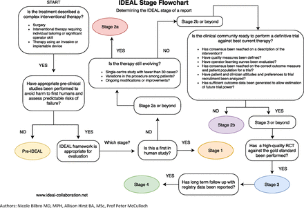
This review revealed fluorescence imaging to enhance the identification of target structures, relative to naked-eye examination or standard white-light imaging, for all five procedures that we evaluated and in the majority of previously published studies, including RCTs conducted on cholangiography and parathyroid identification. Especially for hepatobiliary surgery and (para)thyroidectomy, comparative studies have also documented the advantages of employing fluorescence imaging to enhance the visualisation of anatomical structures and, in doing so, improve short-term and long-term postoperative outcomes. Our IDEAL framework analysis indicates that currently published evidence for fluorescence cholangiography and parathyroid identification is in stage 3, whereas for hepatic segmentation, it is at stage 2b and ready to proceed to well-designed RCTs. For lung segmentation and ureterography, using fluorescence imaging requires further prospective, collaborative, cohort studies to reach final consensus on the best methods and outcome measures to use before proceeding to an RCT (stage 2a), especially for ureterography, for which newer intravenously injected fluorophores that collect in urine may totally alter the standard route of dye administration.

Fluorescence cholangiography using ICG was originally developed for open surgery.37 38 Since fluorescence imaging by intravenous injection enables radiation-free and incisionless intraoperative cholangiography, the best indication for this technique is LC, the most common surgical procedure performed worldwide (750 000–1 000 000 cases annually in the USA alone39). In fact, after the first clinical application of fluorescence cholangiography for LC in 2009,40 its safety and efficacy have been reported for more than 3000 cases, mainly during laparoscopic or robot-assisted surgery. Recently, the superiority of fluorescence cholangiography over standard white-light imaging at delineating the extrahepatic biliary anatomy was demonstrated in an international, multicentre RCT,8 leading to the recommendation that this technique be used as ‘an adjunct to white light alone’ to limit the risk and severity of bile duct injury during LC in multisociety practice guidelines published in 2020.39 Although it may be statistically difficult to orchestrate an RCT large enough to identify a statistically significant impact of fluorescence cholangiography, in terms of reducing the incidence of such a rare but potentially catastrophic complication as bile duct injury during LC (0.15%–0.36%39), further large studies using registries and other similar databases41 should allow for rare favourable and unfavourable events to be revealed as well as educational and cost-efficiencies, and the limitations of fluorescence cholangiography where conventional radiographic cholangiography is needed, moving the use of fluorescence imaging for this purpose to the highest development stage (IDEAL stage 4). Already documented have been reductions in operative times.42 43

Injecting ICG into portal branches (positive-staining technique21 22) to achieve hepatic segmentation is a reappraisal of the conventional dye-staining technique.44 Similarly, administering ICG intravenously following closure of the portal pedicles (negative-staining technique21) builds on the Glissonian approach.45 Since fluorescence imaging enables surgeons to visualise targeted hepatic segments with higher signal-to-background ratios than the naked-eye examinations and standard white-light imaging used with conventional techniques,46 47 the number of publications on both of these newer techniques has increased dramatically in the past 3 years. The expected role of fluorescence-guided hepatic segmentation is to aid the accurate identification of intersegmental planes during hepatectomy procedures which, in turn, should enhance surgical outcomes like operation time, the incidence of complications associated with bile leaks and ischaemia in the remnant liver and tumour-free surgical margins, as suggested by previous prospective studies.20 23–26 The negative-staining technique is simple to perform, while the feasibility of the positive-staining technique seems to be improved by employing preoperative puncture before insufflation48 and intracorporal puncture by robotic manipulation.49 Thus, the IDEAL framework recommends multicentre RCTs to compare the efficacy of anatomic hepatectomies performed with vs without ICG fluorescence imaging for hepatic segmentation.

Interestingly, two approaches—intravenously administering ICG after interrupting blood flow into tumour-bearing lung segments50 and directly injecting ICG into the target bronchi51—can be used for fluorescence-guided lung segmentation, as for hepatic segmentation. The main objective of performing anatomical resections based on lung segmentation is to balance cancer curability and the respiratory function of the remnant lung, reducing the incidence of postoperative complications associated with ischaemia and air leaks.27 Since the former technique—visualising targeted lung segments as non-fluorescing regions—is simple and well established, it is currently recommended that the efficacy of using fluorescence imaging for lung segmentation is compared, in terms of patient outcomes, with lung resections performed using the conventional inflation-deflation method in prospective multicentre studies and RCTs. Other issues usually resolved prior to achieving IDEAL stage 2b have yet to be resolved, however—like which outcomes to use and a consensus definition of the procedure—which is why we still consider this procedure in IDEAL stage 2a, despite the call for RCTs. As for the intrabronchial injection of ICG, although its use with fluorescence imaging for lung segmentation has the potential to improve the signal-to-background ratio, which could decrease surgical times relative to injecting ICG intravenously, additional studies are needed to create consensus on the timing and dose of intravenously injected ICG prior to its adoption in larger prospective studies.

The purpose of using fluorescence imaging for intraoperative ureterography is to prevent injuries to ureters that usually are hidden behind overlying retroperitoneal tissues. Such ureteral injury can occur during a variety of surgical procedures to treat colorectal, gynaecological and urological diseases. Urinary tract injection of ICG has been used as the main approach for fluorescence imaging, although it requires catheterisation into the urinary system.52 53 Another approach is to use the urinary excretion of MB following its intravenous injection, which can omit urinary access but currently requires prototype camera systems fine tuned to detect fluorescence signals in the 680 nm emission range.54 55 Multispectral fluorescence systems are currently being developed commercially, and this will allow for more widespread utilisation of MB fluorescence imaging for ureters in the near future. For further clinical installation of this technique, it is essential to clarify the advantages and disadvantages of each approach based on the patient’s condition and the specific surgical procedure being performed. On the other hand, novel fluorophores optimised for use in ureterography are also being developed28 29; and, while promising outcomes have been reported in early-phase clinical trials, the potential for future development of fluorescence imaging to visualise ureters ultimately largely depends on its cost-benefit performance in preventing urinary tract injuries.

Fluorescence imaging to identify parathyroid glands is unique, in that it relies solely on the glands’ autofluorescence, which makes the procedure easier and safer than other imaging techniques that require the administration of a fluorogenic agent. In contrast, fluorescence imaging using ICG enables the assessment of blood perfusion, which may enhance the prediction and prevention of postoperative hypocalcaemia. In this field, further large studies need to both identify conditions requiring the use of ICG and evaluate the learning curve and cost efficiency of both fluorescence techniques. Although the mechanism behind ICG uptake into glandular tissues remains unclear, this phenomenon also can be used for near-infrared fluorescence imaging of the thyroid and adrenal grand.56 In this review, parathyroid identification was selected as a representative use of fluorescence imaging in endocrine surgery because it was often difficult with other endocrine surgery procedures to determine whether the aim of fluorescence imaging was to identify the gland or the tumour.

Advantages of using the IDEAL framework to overview the development status of fluorescence imaging are not only to recommend the optimal design of future studies in each research area but also to promote information sharing beyond surgical specialties, which should lead to further evolution and dissemination of the technique for intraoperative anatomy visualisation. In this regard, liver surgeons directly communicating their experiences and results using both positive and negative staining techniques for hepatic segmentation have already aided thoracic surgeons in the clinical development of fluorescence-guided lung segmentation. The process of achieving consensus on the intravenous versus intrabiliary injection of ICG will similarly serve as a reference for the future development of ureterography with fluorophores, whether given intravenously or via a catheter directly into ureters. On the other hand, ureteral imaging using urinary stents with a fluorescence resin30 may accelerate the clinical application of fluorescing biliary stents.

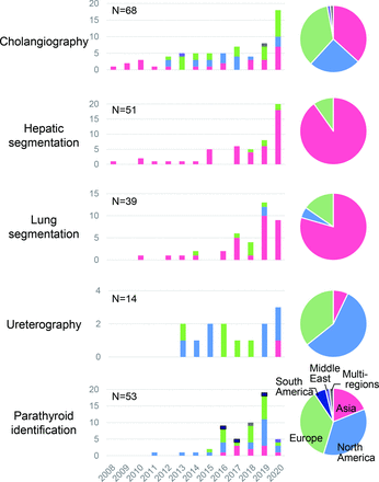
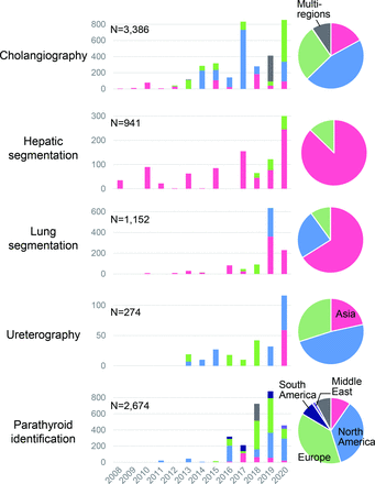
Our analysis has also revealed regional trends in publications and reported cases which, in turn, can elucidate the current dissemination status of these various surgical procedures enabled by fluorescence imaging as well as how commonly each fluorescence imaging technique is being used. For example, fluorescence cholangiography and parathyroid identification (both rated IDEAL stage 3) were introduced in specific regions in the world. However, recent reports come almost equally from the USA, Europe and Asia. In contrast, publications on hepatic segmentation remain largely limited to Asian countries because, even prior to the introduction of fluorescence imaging, anatomic segmentectomy of the liver mainly was advocated for in Asian rather than Western countries. The dissemination of information on fluorescence imaging techniques using the latest near-infrared imaging systems should further contribute to widening the applications of these specialised surgical procedures all over the world.

In conclusion, the current development status of intraoperative fluorescence imaging for visualising anatomy during the five surgical procedures analysed herein lies between IDEAL stage 2a and IDEAL stage 3, with development most advanced for hepatobiliary tract and parathyroid visualisation, intermediate for hepatic segmentation and least advanced for lung segmentation and visualising ureters. For each of these procedures, overviewing the current accumulation of evidence employing the IDEAL framework provides guidance regarding which kinds of subsequent study are needed for each fluorescence imaging technique to aid its development into an essential intraoperative navigational tool.